Certification
AMED CERTIFIED MICROSCOPE USER REQUIREMENTS:
- AMED Member for 2 years
- Passed 2 levels of microscope skills test
- Approved by the AMED Board/Certification Committee
- Attend AMED Annual meeting Certification Ceremony
LEVEL ONE
Exercise A) Identification of the Microscope Parts
The candidate will be asked to identify the following seven parts of the microscope and briefly describe their function:
- Oculars
- Ocular fine focus ring adjustment
- Beam Splitter
- Camera Mount
- Magnification adjustment mechanism
- Fine focus adjustment of the scope or Variable focus adjustment knob
- Main focal lens
Exercise B) Doctor Seating and Ergonomics
The candidate must be able to be seated and adjust their seating position. Thighs should be between 90 degrees to 110 degrees to the floor. The spine should be straight with no forward lean. Head should be in a neutral, relaxed, and balanced position over the shoulders. The eyes should be looking forward and the line of sight parallel to the floor. Arms should rest on the arm supports of the operator chair or be supported and braced against the operator’s sides.
Exercise C) Parfocaling the Microscope
The candidate must demonstrate without assistance or coaching the ability to parfocal the microscope, either the operator specific parfocaling technique or the video-based technique. If the candidate elects to do the operator specific technique the examiner will assist the candidate in video parfocaling the scope in order to observe the examination on the monitor (candidate must video parfocal unassisted to pass level 2 so if candidate is planning to test out of both levels at one seating, this should be done at this time).
Exercise D) Dialing in the Object Tooth
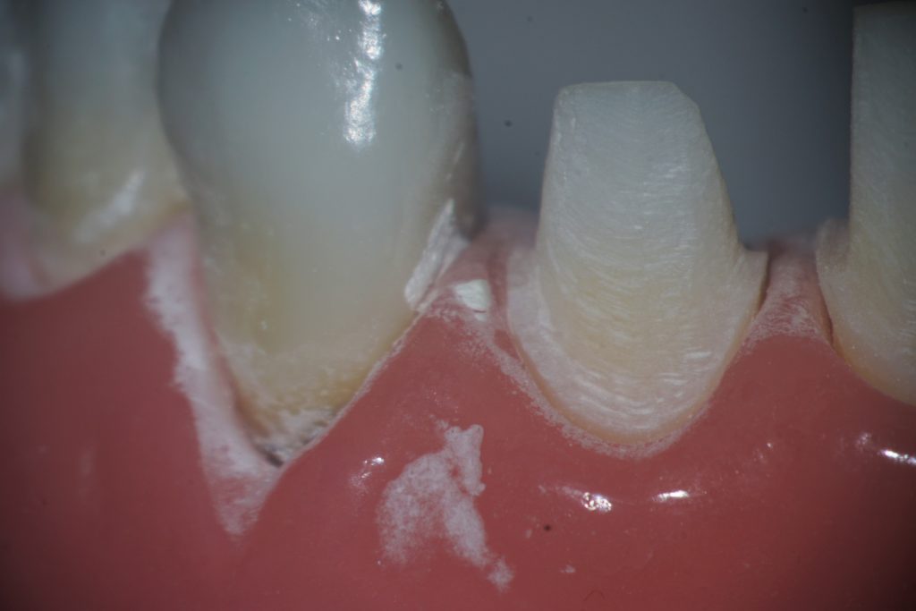
The candidate will be evaluated in bringing the object tooth into focus at a field of view of up to 4 teeth. The candidate may begin at a lower magnification but must complete the exercise in the area of 8X magnification. The candidate must be able to bring tooth #8 into view, use a mirror to visualize the lingual surface and examine the tooth with an explorer. The candidate will then turn to either tooth #3 or tooth #14 (candidate choice) and repeat this exercise.
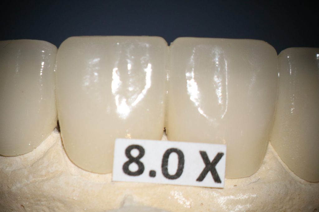
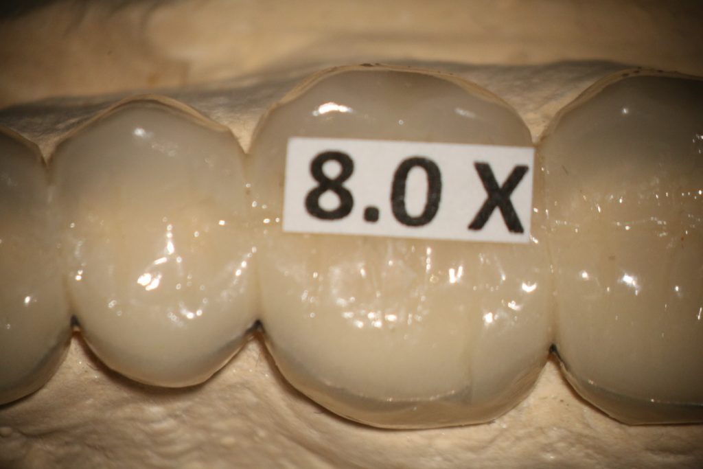
Exercise E) Crown Prep #8
The candidate is permitted to begin at a low magnification but must complete the exercise at a field of view of 4 teeth with an estimated magnification of 8X. The candidate will prepare the facial of tooth #8 then proceed to the mesial and distal proximal reductions. The candidate is not permitted to impact or damage the simulated gingival tissue. The candidate is allowed to utilize a proximal barrier to protect the adjacent teeth from iatrogenic injury. If the adjacent teeth are damaged to the extent that polishing will correct the defect, the candidate will be allowed to pass this section. If the candidate impacts the adjacent tooth/teeth to the extent the tooth would require restoration, the candidate will fail this exercise.
The candidate will proceed to the lingual and prepare the tooth with indirect vision. The margins must connect and be contiguous around the tooth. At least one of the two maxillary procedures must be in focus and centered on video monitor for 50% of five minutes of the procedure.
Exercise F) Crown Prep
Either #3 or #14 – Candidate Choice
The same criteria are applicable as in the anterior tooth Exercise E. No damage to the simulated gingival tissue and no damage to the adjacent teeth that would require restoration.
In Exercises E and F the Examiners are looking for how well the candidate can control the operating instruments and the candidate’s ability to obtain good focus on the object tooth and remain in focus centered in the field of view. The candidate must also demonstrate the ability to operate at a level of magnification 8X with fine motor control and confidence.
LEVEL TWO
The candidate is expected to perform, without coaching or assistance, the following:
Exercise A) Doctor Seating and Ergonomics
Same as level 1.
Exercise B) Parfocaling the Microscope and Video
Candidate must be able to parfocal the microscope to video.
Exercise C) Dialing in the Object Tooth
Same as level 1, substituting #24 and #19 or 30.
Exercise D) Full Crown Prep Anterior Tooth #24
All the same criteria apply from the Level I examination. The crown prep must be completed to the point the tooth is ready for the impression. The margins must be finished and polished. The candidate may begin the exercise at a lower magnification, but the candidate must demonstrate control and mastery with a single tooth operating field of view. The margins must be refined and polished at 14X. The candidate must use the mirror with indirect vision at 14X.
Exercise E) Posterior Tooth Preparation. (Candidate option: MOD Onlay Prep or Full Crown Prep #19 or 30)
The candidate may select the procedure. The same criteria apply to these procedures as to the procedures from Level I preparations.
Keep working field in focus and centered with indirect vision for 70% of a ten-minute portion of the procedure, utilizing at least 8x magnification for at least one minute of the procedure.
The crown prep or the onlay prep must be completed and ready for impression. The margins must be finished and polished at 14X and a mirror must be used during the procedure at the 14X level.

FREQUENTLY ASKED QUESTIONS
Do I have to be an AMED Member to receive certification?
Yes, 2 years of consecutive membership are required to receive certification.
Do I have to be a member for two years before I take the certification exam?
No, you can take the exam the first year if you want.
Do I have to take both levels of the exam at once?
No, you can take each level at separate meetings or both at one meeting.
Where is the exam offered?
The exam is only offered at the AMED Annual Meeting.
Do I have to attend the meeting to receive the certification?
Yes, attending the awards ceremony at AMED’s Annual Meeting is required to receive the award.
Can I bring my own instruments for the exam?
Yes.
What is the Cost?
For One Level: $395
For Two Levels: $695 if you take both levels at the same time.